Angioplasty
Information and Appointment Line
e-Services

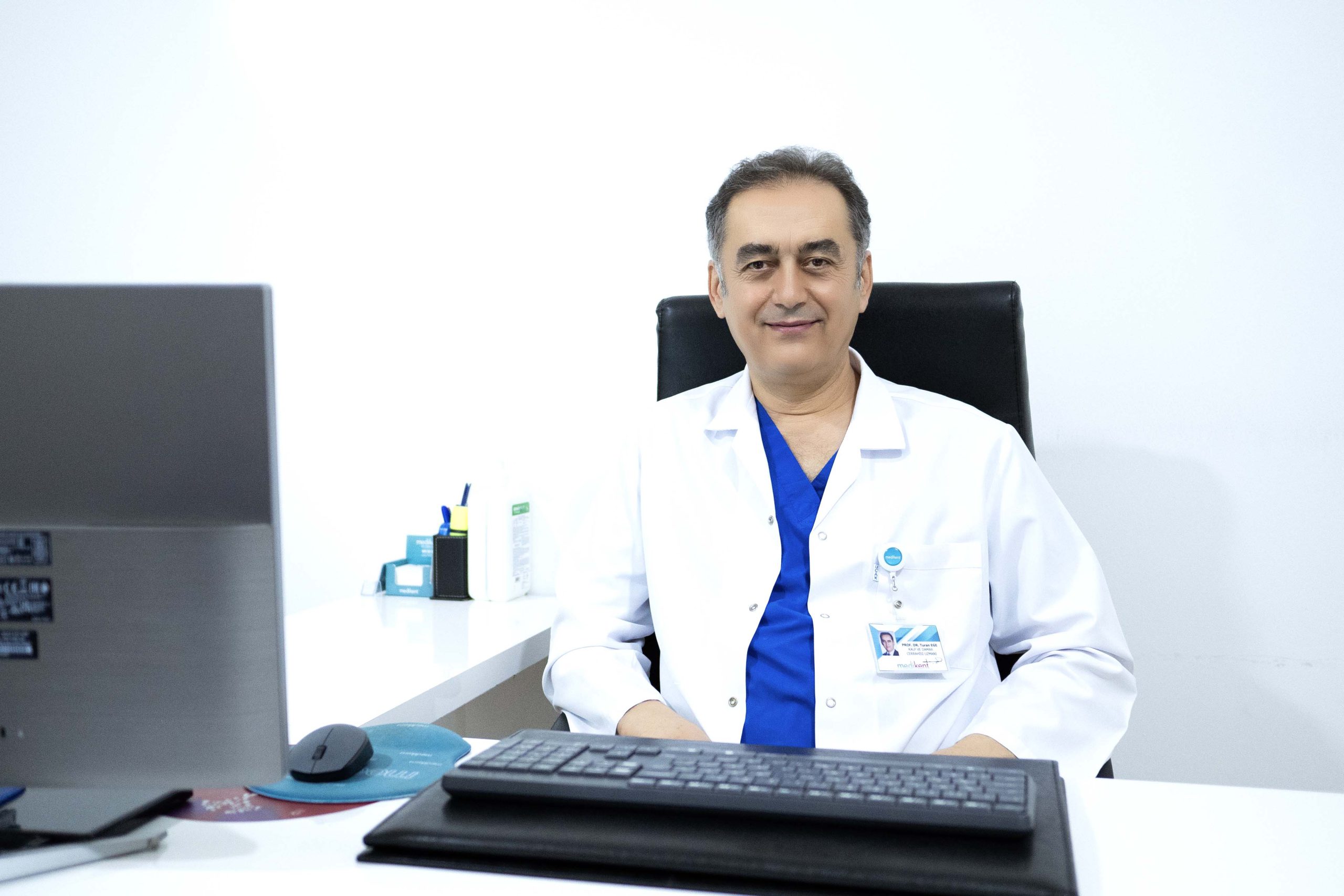
Cardiovascular Surgery Prof. Dr. Turan EGE

Cardiology SpecialistProf. Dr. Bilal GEYİK

Cardiology SpecialistSpecialist Dr. Haydar Başar CENGİZ
Angioplasty
Coronary artery disease is the narrowing of the arteries that feed the heart as a result of the development of atherosclerosis (arteriosclerosis). The presence of risk factors such as smoking, hypertension, especially fat around the navel and diabetes causes the formation of atherosclerosis (endothelial dysfunction). If not recognized in time and the necessary measures are not taken, blocked blood vessels can lead to heart attack (myocardial infarction) and fatal rhythm disturbances. Coronary artery disease is the leading cause of death and labor loss in our country and the world. Genetic predisposition is very important in the development of coronary artery disease, as it is in many diseases, and individuals with a family history of early heart attack are particularly at risk. Irregular and high-fat diets lead to an increase in cholesterol and other harmful blood fats and predispose to the development of atherosclerosis. People who lead stressful lives, do not exercise and choose a sedentary lifestyle are at risk of developing coronary artery disease.
While it may progress for years without any symptoms, new onset of fatigue, chest, back and arm pain that comes with exertion and restricts exertion, and new onset of shortness of breath related to exertion are common symptoms. In addition, the first sign of coronary artery disease can be a sudden heart attack or, in less fortunate patients, sudden death.
If coronary artery disease is suspected, certain tests are ordered by specialists to diagnose the disease. An electrocardiogram (ECG) is an absolute must in every patient. This test provides information about the heart’s beating pattern and the presence of a previous heart attack. The fact that it can be taken at the time of the patient’s complaint increases its diagnostic value. An ECG taken in the absence of symptoms may be normal, so a normal ECG does not rule out the presence of coronary artery disease. If necessary, a stress ECG (running test) may be ordered to assess whether blood supply is impaired during exercise. Myocardial perfusion scintigraphy is a more sensitive, but more expensive and radiation-risk-carrying method than a stress test that evaluates the blood supply to the heart using nuclear medicine methods. Echocardiography (ultrasound of the heart) provides detailed information about the contraction of the heart and the condition of the heart valves. The gold standard in the diagnosis of coronary artery disease is undoubtedly coronary angiography.
A cannula is inserted into a large artery in the arm or leg and a dye is injected through the cannula into the small arteries (coronary arteries) that supply the heart. This procedure helps to visualize the coronary vessels on the X-rays and to determine how much stenosis is present in which areas.
Angiography is the most definitive test method that shows whether the patient has coronary artery disease. Angiography can determine the extent to which the coronary arteries are narrowed or blocked by atherosclerosis. If the angiography reveals a blockage or narrowing of the vessel, it is decided whether it should be treated with a stent or surgery.
Coronary angiography should be recommended in the presence of chest pain suggestive of cardiovascular disease, in patients who have had a heart attack, in patients with risk factors and abnormal results of tests suggestive of vascular disease, in patients who develop heart failure or severe rhythm disturbances without an explanatory reason. In addition, coronary angiography should be performed in patients who will undergo heart or vascular surgery for reasons other than heart disease, such as valvular disease or vasodilation, and in patients with a history of stenting or bypass and recurrent complaints. Coronary angiography performed under emergency conditions during a heart attack, followed by vascular opening procedures such as balloon/stenting, is life-saving.
The preparation phase of angiography takes 5 minutes and the procedure takes 5-10 minutes, totaling 10-15 minutes. It may take up to 10-15 minutes to apply pressure on the entered vein.
In this rare type of cardiomyopathy, the muscle in the lower right heart chamber (right ventricle) is replaced by scar tissue, which can lead to heart rhythm problems. It’s often caused by genetic mutations.
Medikent Angioplasty Your Heart is Safe with Us!
As Medikent Angiography Center, we focus on your heart health and perform angiography procedures with the most modern methods. With our experienced cardiologists and state-of-the-art equipment, we are here to protect your heart’s health at the highest level. For a healthy life, contact us now and book your angioplasty appointment!

